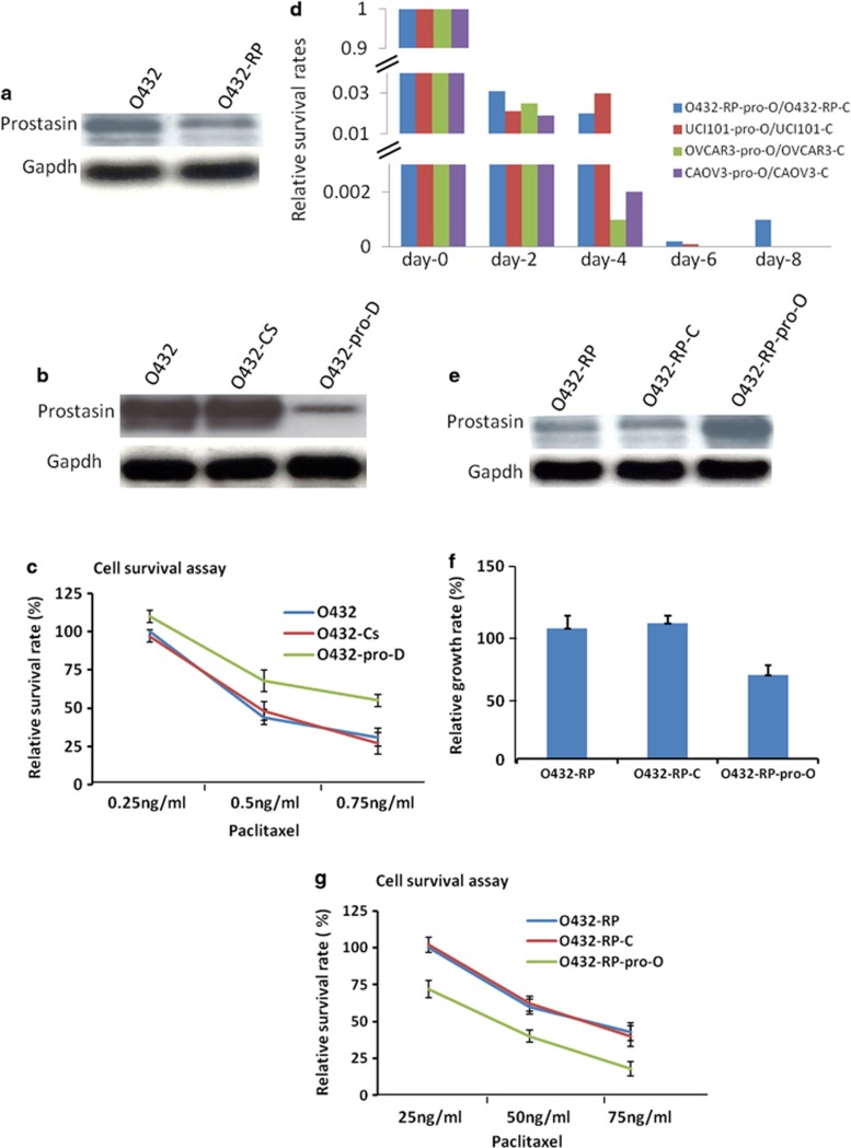
Figure 2.
Prostasin has important roles in chemoresistance and cell death in cell culture model. (a) Prostasin decreased in paclitaxel-resistance cancer cell line. O432: ovarian cancer cell line Ovca432 (sensitive to paclitaxel); O432-RP: paclitaxel resistance cell line generated from Ovca432. The prostasin protein levels in O432 and O432-RP cells are shown in immunoblots with specific antibodies. (b) Prostasin siRNAs transfection reduced prostasin. O432-pro-D cells (transfected with prostasin siRNA) express lower prostasin, compared with control cells of O432 (transfected with reagent only) and O432-Cs (transfected with no-targeting siRNA). The prostasin protein levels are shown in immunoblots with specific antibodies. (c) Downregulation of prostasin in O432 cells resulted in increase of chemoresistant activity. Cells were treated with paclitaxel at different concentrations for 24 h (starting 48 h after siRNAs transfection) and cultured with normal medium for an additional 7 to 10 days before cell survival was assayed. Relative cell survival rates of each cell line are shown. (d) Overexpression of prostasin greatly induces cell death in ovarian cancer cells. The cell survival rates are shown after forced overexpression of prostasin in several cell lines from day-0 to day-8, respectively. (e) Prostasin cDNA transfection resulted in overexpression of prostasin in chemoresistant O432-RP cells. O432-RP-pro-O cells (transfected with prostasin cDNA) express higher prostasin compared with control cells O432-RP and O432-RP-C (transfected with control vector). The prostasin protein levels are shown in immunoblots with specific antibodies. (f) Forced overexpression of prostasin represses growth of chemoresistant cells. Relative cell growth rates are shown for O432-RP-pro-O and control cells O432-RP and O432-RP-C. (g) Forced overexpression of prostasin in O432-RP cells re-sensitizes chemoresistant cells. Cells were plated at about 10–20% confluence and treated with paclitaxel at different concentrations for 24 h, cultured with normal medium for additional 7 to 10 days, then assayed for cell survival. Relative survival rates of cell lines are shown

