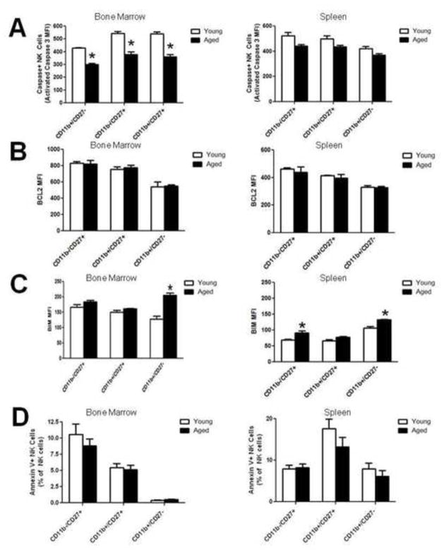
Figure 5. Analysis of the apoptotic potential of resting NK cells from young and aged mice.
(A) Freshly isolated bone marrow and spleen cells were stained intracellularly for caspace-3. Median Fluoresence Intensity (MFI) of expression for each NK cell subset, (n=5). (B) Freshly isolated bone marrow and spleen cells were stained intracellularly for Bcl-2. MFI of expression for each NK cell subset, (n=5). (C) Freshly isolated bone marrow and spleen cells were stained intracellularly for BIM. MFI of expression for each NK cell subset, (n=5). (D) Annexin staining for each NK subset for bone marrow and spleen cells incubated for 10 hrs without growth factors at 37°C, (n=10). Asterisks indicate statistical significant difference between the two age groups, t-test, p < 0.05.

