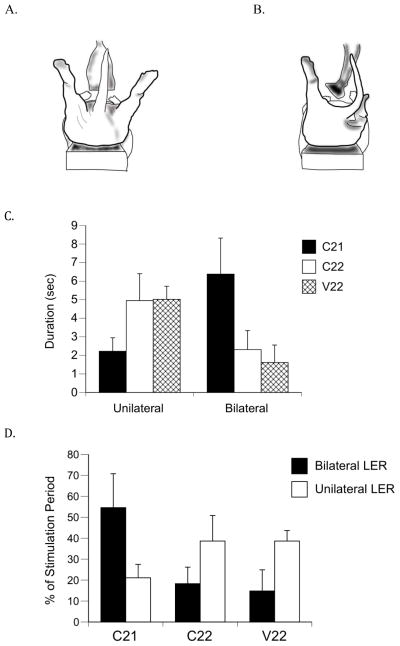
Figure 2.
Unilateral and bilateral expressions of the LER. (A) Illustration of the bilateral and (B) unilateral LERs expressed during anogenital stimulation. (C) Absolute duration in seconds of unilateral and bilateral LERs and (D) mean percentages of unilateral and bilateral LER durations during the 15-sec stimulation period. C21 subjects showed a significantly longer absolute bilateral LER duration and significantly longer bilateral LERs as a percentage of stimulation time compared to subjects in the control groups (C22 and V22). Bars show means; vertical lines depict SEM. There were 8 subjects each in the C21 and V22 groups, and 6 subjects in the C22 group that showed the LER.

