Figure 6.
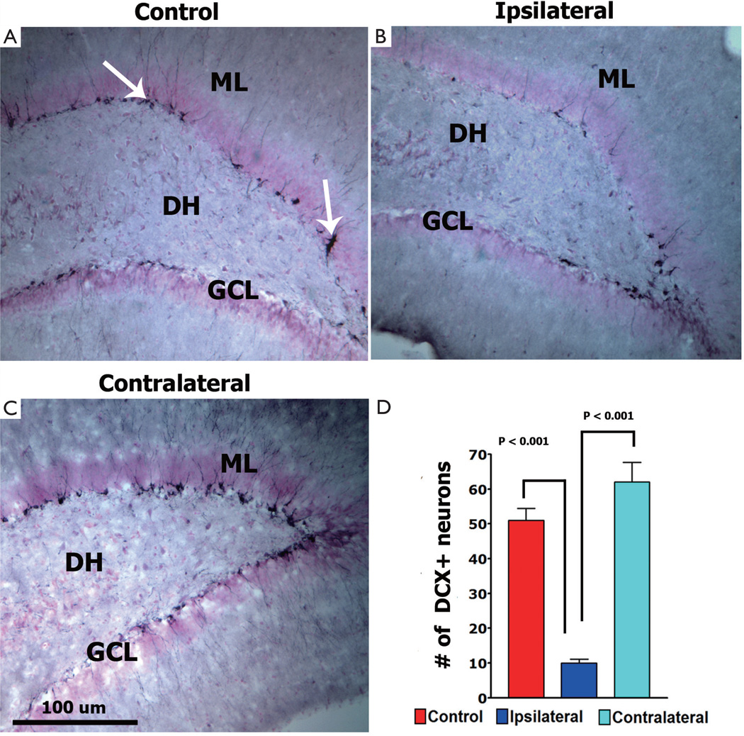
Digital photomicrographs of early neurogenesis in the dentate gyrus as analyzed by DCX immunostaining (A–C). The ipsilateral hippocampus (B) showed reduced (80%, P<0.001) numbers of DCX-positive newly born neurons while the contralateral hippocampus showed increased (20%) numbers of these cells compared to unirradiated controls. Bar chart (D) shows the quantification of DCX-positive cells (arrows) in the unilaterally irradiated (10 Gy) brain

