
Fig. 2.
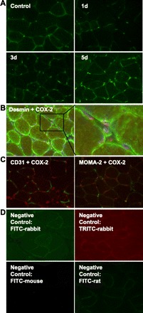
COX-2 is expressed in muscle during compensatory hypertrophy. A: COX-2 expression in plantaris was visualized by immunofluorescence in control muscle and at 1, 3, and 5 days postablation. B: localization of COX-2 expression at 5 days postablation was determined using immunofluorescence for COX-2 (red) and desmin (green). Nuclei were counterstained with 4′,6 diamidino-2-phenylindole (blue). COX-2 fluorescence was most often associated with nuclei within or near desmin-positive cells. C: immunofluorescence for COX-2 (red) and CD31 (green, left) or MOMA-2 (green, right) at 5 days postablation. COX-2 exhibited little colocalization with either the endothelial cell marker CD31 or the monocyte/macrophage marker MOMA-2. D: representative images of negative controls for muscles at 5 days postablation. Negative controls at other time points were similar. All images in Fig. 2 are representative of at least 4 muscles per condition.