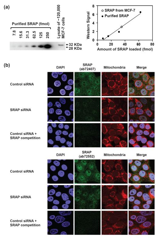
Figure 1. SRAP expression level and localization in MCF-7 cells.
(a) SRAP quantitative western blot. Left: Lanes 1-6, purified SRAP (13-236) protein; lane 7, lysate from ~120,000 MCF-7 cells. SRAP was probed with Abcam antibody ab72552. Right: plot of western signal versus amount of SRAP. Open circle, signal from cell lysate. (b) Immunofluorescence analysis of SRAP in MCF-7 cells. SRAP was probed with Abcam antibodies ab72407 (top panels) or ab72552 (bottom panels). The signal specificity was confirmed by knocking down SRAP with pool 2 SRA siRNA (middle rows) as well as by competition with recombinant SRAP (13-236) protein (bottom rows).

