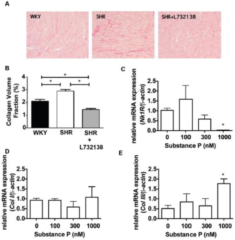
FIGURE 4.

A. Collagen was stained with picrosirius red in 24-week old left ventricular tissue slices from WKY, SHR, and SHR treated with L732138. B. Collagen volume fraction was quantified from the picrosirius staining (A). n=8 for each group, WKY (black bar), SHR (white bar), and SHR treated with L732138 (grey bar). *p<0.05, 1-way ANOVA with a Tukey post-test. C. Nk-1 receptor mRNA was measured and expression was normalized to β-actin in cardiac fibroblasts incubated with increasing concentrations of substance P for 24 hours. Relative fold expression was calculated using the ΔΔCt method. *p<0.05, 1-way ANOVA with a Tukey post-test. D-E. Collagen I (D) and Collagen III (E) mRNA was measured in substance P treated cardiac fibroblasts after 24-hours. Fold expression was determined using the ΔΔCt method. *p<0.05, 1-way ANOVA with a Tukey post-test.
