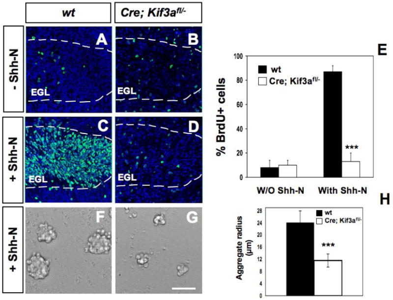
Fig. 6. Abnormal Shh-induced proliferation of GCPs in cerebellar cultures from conditional Kif3a mutants.

BrdU staining on cerebella sagittal slices from P3 wild type (A, C) and mutant mice (B, D) cultured with no stimulus (A-B) or 3 μg/ml Shh-N (C-D). BrdU staining (green) was counterstained with DAPI (blue) and visualized using confocal microscopy. (E) Quantification of the percentage of BrdU+ and DAPI+ nuclei in wild type and conditional Kif3a mutant slices with and without Shh-N. (F-G) Proliferation of isolated GCPs in aggregates obtained from P1 Kif3a mutants (G) compared to controls (F) with Shh-N for 48h. (H) Quantification of aggregate radius in wild type and conditional Kif3a mutants after 48h. Data from three mice per group were pooled for statistical analysis with Student’s t-test. ***: p<0.001. Scale bar: 100 μm.
