
Fig.7. Abnormal proliferation of GCPs in conditional Smo mutant and conditional Kif3a and Smo double mutant mice.
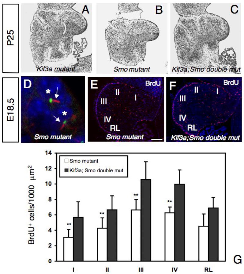
(A-C) Cresyl violet staining of sagittal sections of cerebellum from conditional Kif3a mutant (A), conditional Smo mutant (B), and conditional Kif3a and Smo double mutant cerebellum at P25. Rostral is to the right. (D) Double immunostaining with an anti-acetylated α-tubulin antibody (red) and an anti-γ-tubulin antibody (green) on sections of conditional Smo mutant at E18.5. In conditional Smo mutants, most GCPs extend a primary cilium (arrow) from a basal body. Each nucleus associated with a primary cilium is indicated by an asterisk. (E-F) BrdU staining of a conditional Smo mutant (E) and a conditional Kif3a and Smo double mutant (F) cerebellum at E18.5. Rostral is to the right. (G) Quantification of BrdU+ cells per 1000 μm2 in regions I to IV and in the RL. Note that the number of BrdU+ cells is much smaller in conditional Smo mutants than in conditional Kif3a and Smo double mutants, while no significant difference was observed between conditional Kif3a mutants and conditional Kif3a and Smo double mutants (compare with Fig.4F). The BrdU+ cells were counted from at least three sections from each mouse at comparable mediolateral levels. Data from three mice per group were pooled for statistical analysis with Student’s t-test. **: p<0.01. Scale bar: 230 μm (A-C), 2 μm (D) and 100 μm (E-F).