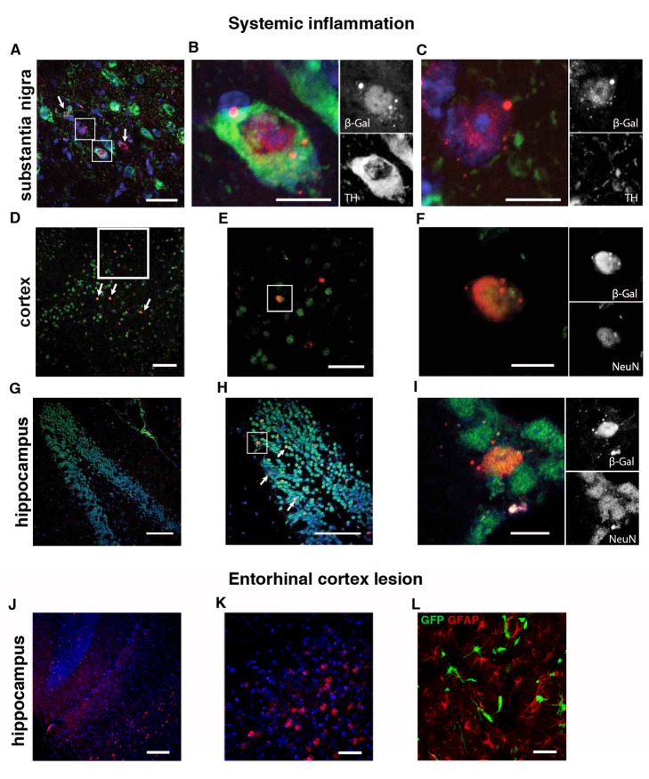
Figure 6. Reporter-gene–positive neurons can be found in multiple areas of the brain.
(A and B) β-galactosidase–positive cells in the SN/VTA that can be TH-positive dopaminergic neurons as well as TH-negative cells with neuronal morphology (C). (D–F) β-galactosidase–positive cells in the cortex are also positive for NeuN. (G–I) Overview of the dentate gyrus in the hippocampus with recombined neurons in the granular cell layer that are positive for the neuronal marker NeuN. (J and K) Recombined neurons after ECL in hippocampal areas CA2 and CA3 and nonneuronal GFAP-negative recombined cells at the lesion site (L). Scale bar, 100 µm (D, G, H, and J), 50 µm (A, E, K, and L), and 10 µm (B, C, F, and I).

