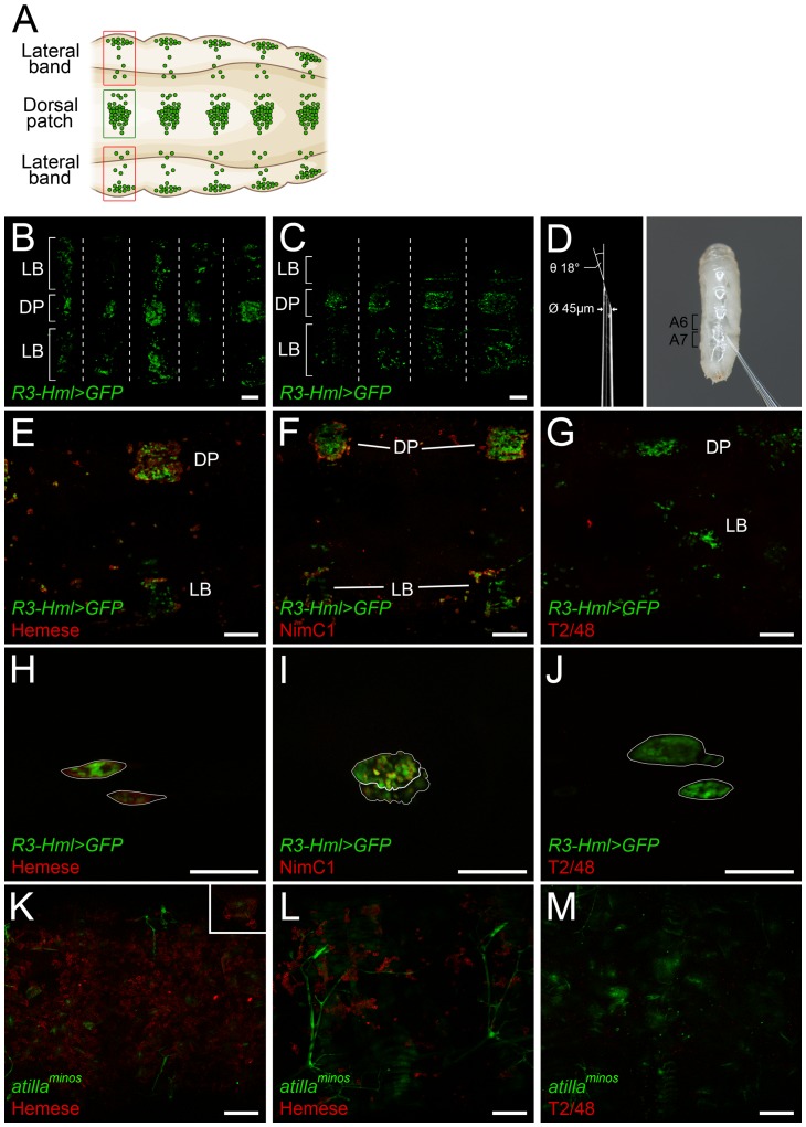
Figure 2. The schematic representation of the larval sessile hematopoietic tissue (A).
The sessile tissue of an immobilized R3-Hml>GFP larva (B), and a mock-injected R3-Hml>GFP larva (C). The dorsal patches are indicated by DP, and the lateral bands are indicated by LB. Segmental borders are marked with dashed lines. The parameters for the sharpened capillaries used for the injection of larvae, and a high-magnification photograph of the injection (D). Staining (red) of the sessile hemocytes (green) in R3-Hml>GFP larva with anti-Hemese (E), anti-NimC1 (F) and T2/48 (G) antibodies. The lymph glands (outlined in white) of R3-Hml>GFP larvae (green) stained in situ with anti-Hemese (H), anti-NimC1 (I) or T2/48 (J) antibodies (red). The sessile hematopoietic tissue of atillaminos; l(3)mbn1 (K) and atillaminos (L) larvae stained for Hemese (red). Negative control staining of atillaminos; l(3)mbn1 larva with T2/48 antibody (M). The lamellocytes (green) of atillaminos; l(3)mbn1 larva in the hematopoietic tissue stained for Hemese (K, insert), indicated by the arrows. All scale bars indicate 50 µm.

