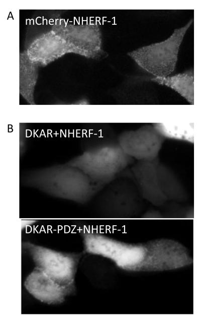
Figure 2.
Fluorescent images of MDCK cells. A. mCherry-tagged NHERF-1 localizes to NHERF complexes at the apical membrane of polarized epithelial cells (e.g. MDCK cells) and this presents in a punctate pattern. B. Untargeted DKAR is present throughout the cytosol and nucleus of the cell (top). Addition of the PDZ-targeting motif to DKAR (DKAR-PDZ) localizes it to the NHERF scaffold in NHERF-1-overexpressing MDCK cells (bottom); as not all cells display relocalization of DKAR-PDZ to the NHERF scaffold, care should be taken to select those that do.

