
Figure 4.
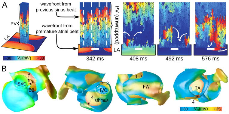
A: Continuous PV re-activation of the LA due to heterogeneous venous conduction and reentry following a single ectopic beat in the LA (with permission from103). The sinus beat propagates heterogeneously along the vein (wrapped and unwrapped views, 342ms). Vein length is 1 cm, and circumference is 2cm, with 30% longitudinal and 65% transverse cellular disconnections. A single premature activation originating somewhere in LA invades PV following the sinus beat (342ms and 408ms), encountering block (408ms) and establishing PV reentry (492ms). This reentry continuously re-excites the LA, serving as “focal source” for LA activations (576ms; propagation entering LA at bottom of image). B: A snapshot of membrane voltage in the human atria at a single time point during AF (different views of the atria are shown), in which numbered arrows (1–4) indicate multiple reentrant wavelets. AF was induced by PV ectopic beats (with permission from64). SVC and IVC–superior and inferior vena cava; BB--Bachman’s bundle; FW–free wall; TA—tricuspid annulus.