
Figure 7.
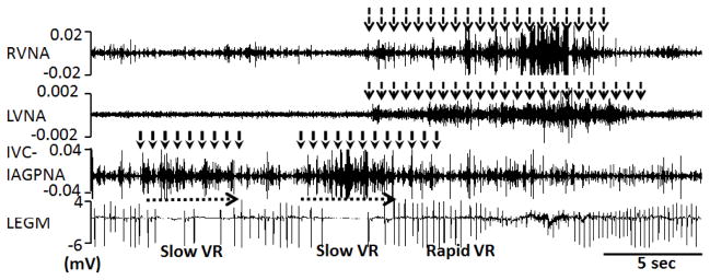
Local control of atrioventricular (AV) node conduction during persistent atrial fibrillation (AF). Slowing of ventricular rate (VR) was associated with inferior vena cava-inferior atrial ganglionated plexus nerve activity (IVC-IAGPNA) without either right vagal nerve activity (RVNA) or left vagal nerve activity (LVNA). Subsequent simultaneous activation of right vagal nerve activity and left vagal nerve activity resulted in a rapid ventricular rate. Because of the presence of abundant sympathetic nerves within the vagus,17 these observations suggest that sympathetic component within the vagal nerves have accelerated the ventricular rate. LEGM is the bipolar local electrogram showing ventricular activation. From Park et al,18 with permission.