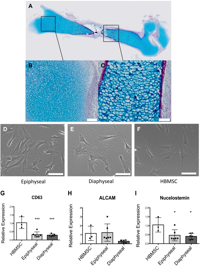
Figure 1. Morphology of foetal femur derived cells in whole femur and post monolayer culture with quantitative expression of stromal antigens and putative stem cell marker by HBMSC, foetal femur epiphyseal and diaphyseal cells.
Foetal femur comprised a proteoglycan anlage (Blue) with a bone collar marked by deposition of collagen (Red) (A). Epiphyseal region contained proliferating chondrocytes (B) while diaphyseal section, comprised hypertrophic chondrocytes (C). Epiphyseal cells (D) and diaphyseal cells (E) adopt similar morphology to HBMSC (F) upon monolayer culture. Expression of CD63 (G), ALCAM/CD166 (H) and Nucleostemin (I) by foetal femur epiphyseal cells and diaphyseal cells was confirmed by RT-qPCR and expression levels compared to human bone marrow stromal cells (set to have expression of one). Results expressed as mean ± SD and n = 3. ***P<0.001 and *P<0.1 calculated using ANOVA. Scale bar = 100 µm.

