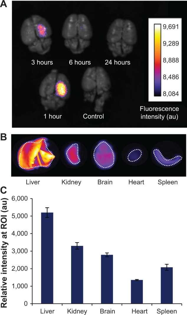
Figure 8.

Ex vivo fluorescence imaging of biodistribution of the nanoparticles in an orthotopic mouse model of PBT.
Notes: (A) Fluorescent signal observed in the brains of four separate mice with PBT (1–24 hours) injected intravenously (via tail vein) with transferrin-coated Prussian blue nanoparticles (MnPB-ATxRd-Tf). A control mouse with PBT was not injected with nanoparticles. (B) Representative ex vivo fluorescence imaging of organ biodistribution of the nanoparticles at 3 hours post-injection. (C) Histograms quantifying the observed fluorescence biodistribution of the nanoparticles 3 hours post-injection. ROIs for intensity measurements are indicated by white dashed lines in (B).
Abbreviations: PBT, pediatric brain tumor; MnPB, manganese-containing Prussian blue; ATxRd, Texas Red-labeled avidin; Tf, transferrin; ROI, region of interest; au, arbitrary units.
