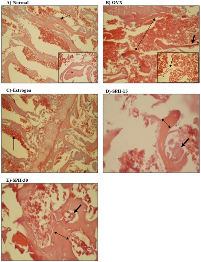
Figure 4. Histopathological assessment of the anti-osteoporosis effect of sophoricoside.
Normal group (A) shows normal bony tissue with intact well-formed dense trabeculae (star in the inner panel) with osteoblastic rimming (arrow) and average intervening bone marrow. OVX group (B) showed scant, disconnected (arrow in inner panel), thin (arrow in the outer panel), and widely separated trabeculae (double-headed arrow in the outer panel) with eroded surface (star in the outer panel). Estrogen treated group (C) showed widely distributed osteoid and osteoblastic rimming (arrow). SPH-15 group (D) showed thick trabeculae (double-headed arrow), more osteoid, and osteoblastic activity (arrow). SPH-30 group (E) showed thick trabeculae (double-headed arrow), more osteoid and osteoblastic activity (arrow).

