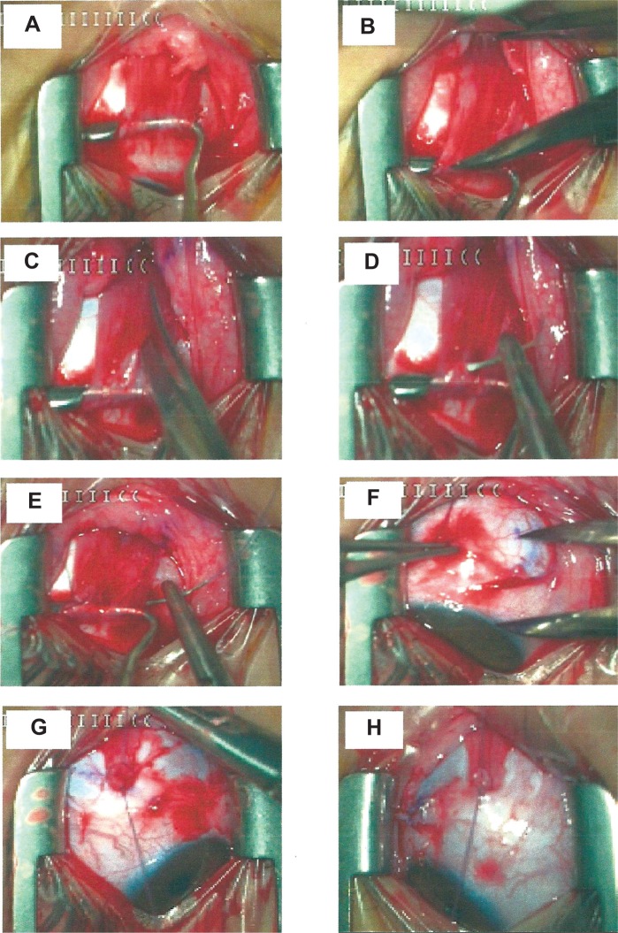
Figure 2.
Steps in Y-split recession.
Notes: (A) The MR is hooked. (B) A 15 mm distance from the insertion is measured. (C) The muscle is split along the 15 mm. (D and E) The upper and lower MR halves are sutured. (F) The minimum distance from the limbus to the new insertion is measured. (G and H) The two muscle halves are sutured at the new insertion points.
Abbreviation: MR, medial rectus.

