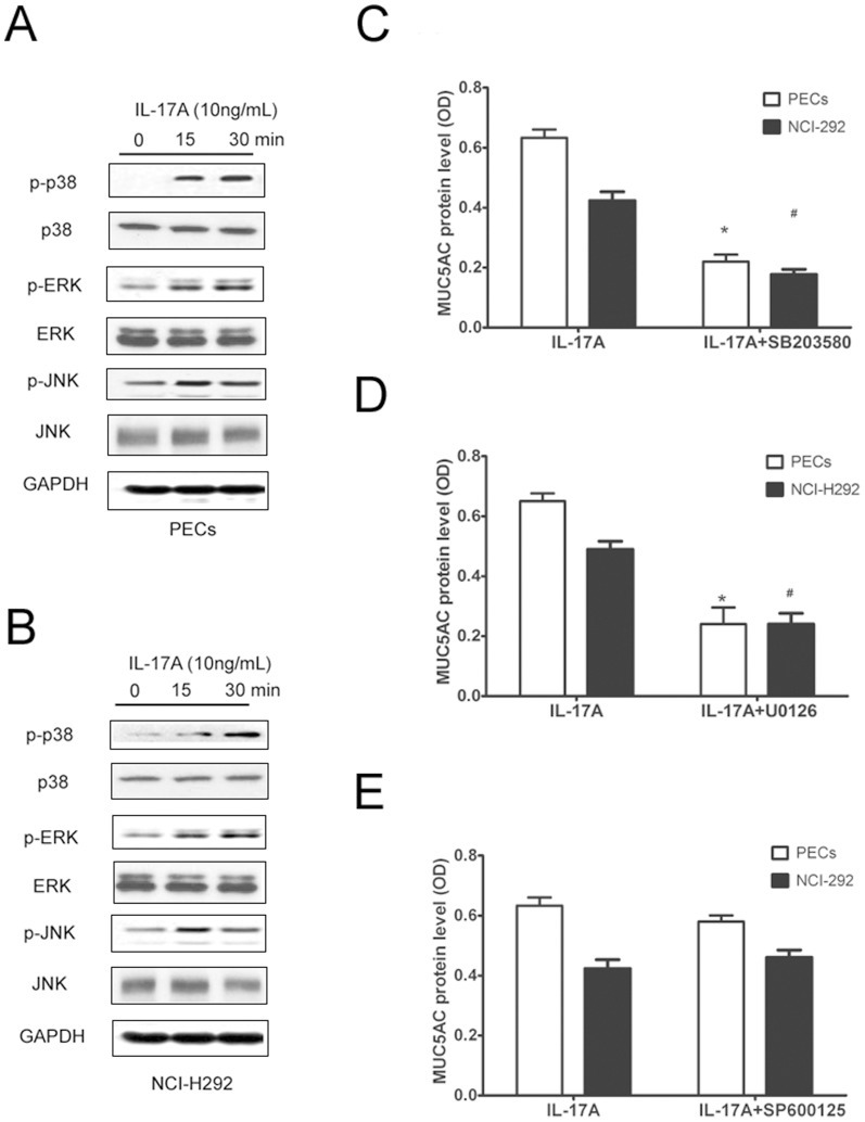
Figure 5. MAPK signaling mediated IL-17A induced MUC5AC in PECs and NCI-H292 cells in vitro.
(A) Representative western blot result of phosphorylated p38, ERK and JNK in PECs after IL-17A stimulation. (B) Representative western blot result of phosphorylated p38, ERK and JNK in NCI-H292 cells after IL-17A stimulation. (C, D) MUC5AC protein level in cultured PECs and NCI-H292 cells after IL-17A stimulation for 24 h in the presence of specific inhibitors of p38, ERK and JNK. The data are expressed the means (SEM) of 3 independent experiments. * p<0.05 when compared with control PECs, # p<0.05 when compared with control NCI-H292 cells.

