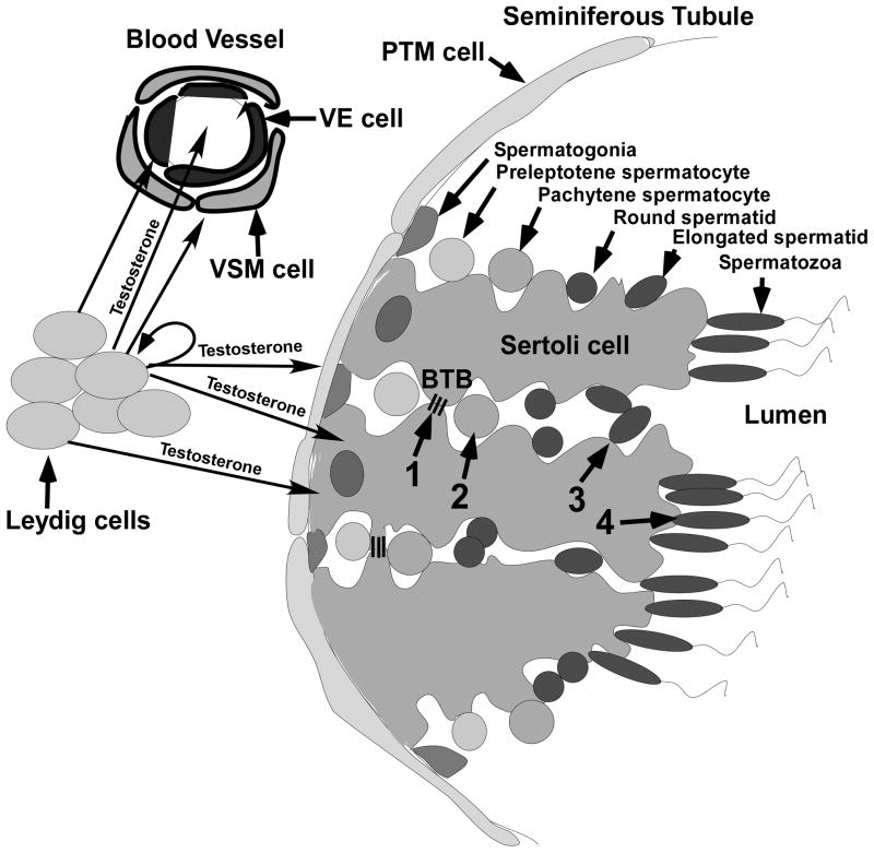
Figure 1.
The anatomy of the testis and the process of spermatogenesis. Leydig cells and blood vessels that are lined by vascular endothelial (VE) and vascular smooth muscle (VSM) cells are localized to the interstitial space between seminiferous tubules. Peritubular myoid (PTM) cells line the outside of the seminiferous tubule. Sertoli cells extend from the PTM cells to the lumen and surround the developing germ cells. The process of spermatogenesis is outlined at the top of the figure where germ cell development is shown progressing toward the lumen from spermatogonia on the basement membrane to the production of spermatozoa. Testosterone produced by the Leydig cells diffuses into the VE and VSM cells as well as into the blood vessels. Testosterone also diffuses from the Leydig cells into PTM, Sertoli cells and Leydig cells. The four critical processes regulated by testosterone (1–4 in the middle of the figure) are indicated: 1) the maintenance of the BTB (represented by 3 lines extending between 2 Sertoli cells), 2) completion of meiosis by spermatocytes, 3) adherence of elongated spermatids to Sertoli cells and 4) the release of mature spermatozoa.

