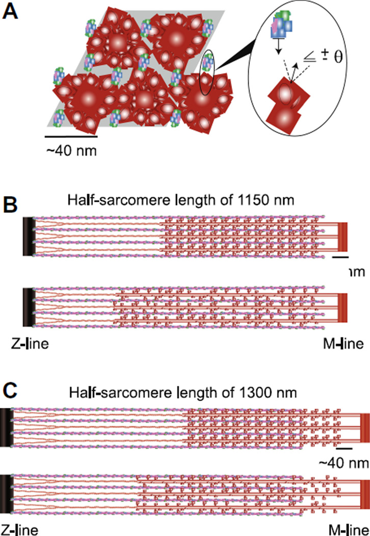
Figure 1. Model geometry and half-sarcomere organization.
A) The shaded parallelogram roughly outlines the cross-sectional view of modeled interactions between four thick-filaments (red) and eight regulated thin-filaments (actin=blue, troponin=green, tropomyosin=magenta) of half-sarcomere length. The expanded inset illustrates one potential myosin-actin cross-bridge interaction between two adjacent filaments. Axial, half-sarcomere illustrations show filament interactions for simulations of B) 1150 nm (=2.3 µm SL) and C) 1300 nm (=2.6 µm SL) for uniform (upper) and random (lower) reductions in myosin content at each SL. Titin is represented as the link between thick-filament backbones and the Z-line.

