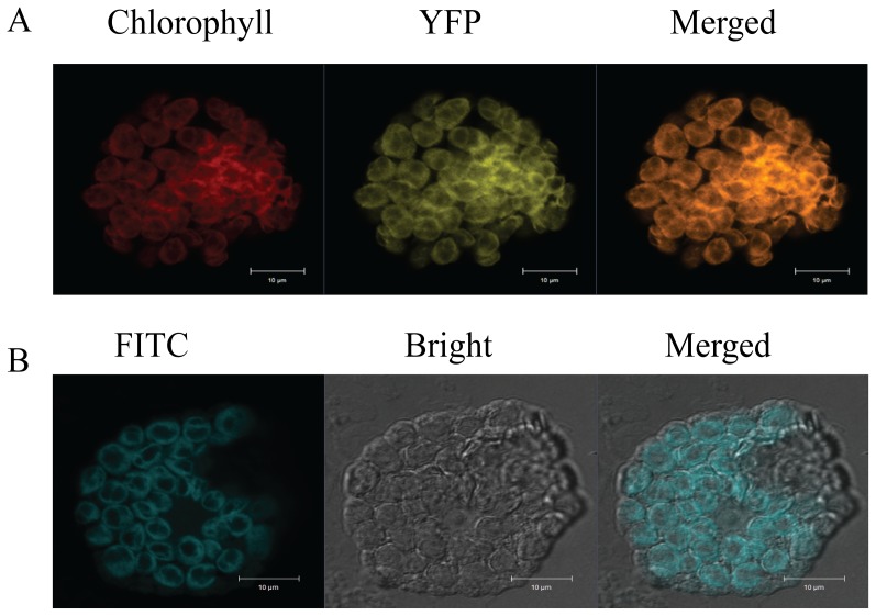
Figure 5. Subcellular localization of HvBT1::YFP.
Subcellular localization of HvBT1 was visualized by Zeiss confocal Laser scanning microscope. A: transient expression of HvBT1::YFP in living protoplasts; chlorophyll autoflorescence (red color), YFP fluorescence (yellow color) and merged image (orange color). B: immunolocalization of HvBT1::YFP was detected using anti-YFP antibody and visualized by the fluorescence of FITC-conjugated antibody. Images represent FITC fluorescence (blue color), bright field (grey image) and the merged image that show the localization of HvBT1::YFP on the chloroplasts membranes.

