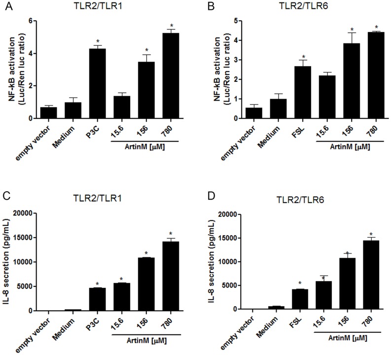
Figure 3. ArtinM induces the activation of TLR2/1- and TLR2/6-expressing cells.
HEK293A cells were transfected with TLR2/1 (A and C) or TLR2/6 (B and D), co-receptors, an NF-κB reporter construct, and a Renilla luciferase reporter plasmid as described for Figure 2. The transfected cells were stimulated with ArtinM (15.6, 156, and 780 nM) at 37°C for 4 h. Medium and cells transfected with an empty vector were used as the negative controls. The positive controls were P3C4 (1 nM) for TLR2/1 activation (A and C) and FSL1 (0.1 nM) for TLR2/6 activation (B and D). The luciferase activity (A and B) was measured as described in the materials and methods. IL-8 levels in the culture supernatants (C and D) were measured by ELISA. Statistical comparisons between the cells incubated with medium and the cells stimulated with ArtinM were performed with a one-way analysis of variance followed by Bonferroni's multiple comparison test. * p<0.05.

