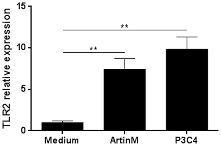
Figure 4. Enhanced TLR2 relative expression by ArtinM-stimulated macrophages.
Peritoneal macrophages from C57BL/6 mice were incubated with ArtinM (39 nM) for 5 h. Medium was used as a negative control and P3C4 (1 µg/mL) was used as a positive control. RNA from macrophages were isolated and used for qRT-PCR as described in Materials and Methods. The results are expressed as the relative expression of TLR2 after quantification using the ΔΔCt method and normalized to β-actin expression. Statistical comparisons between stimulated cells and unstimulated were performed with one-way analysis of variance followed by Bonferroni's multiple comparison test. ** p<0.01.

