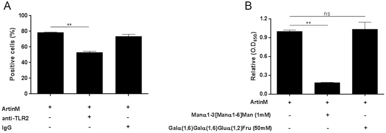
Figure 5. ArtinM binding to TLR2 depends on sugar recognition.
(A and B) Peritoneal macrophages from C57BL/6 mice were incubated with biotinylated ArtinM after pre-incubation with anti-TLR2 antibody or non-specific IgG. ArtinM binding was detected with streptavidin-FITC and analyzed by flow cytometry, as described in materials and methods. Results are expressed as the percentage of cells positive for ArtinM binding (A) and MFI (median fluorescence intensity) (B). (C) The dependence of ArtinM-TLR2 binding on carbohydrate recognition used anti-TLR2 antibody coated onto 96-well microplates (5 µg/mL) to capture TLR2 from a cellular extract of peritoneal macrophages. Biotinylated ArtinM (40 µg/mL), previously incubated with the indicated concentrations of Manα1-3 [Manα1-6] Man or Galα(1,6)Galα(1,6)Gluα(1,2)Fru, was added to the wells. After washing, ArtinM binding was detected by neutravidin-AP, and signal was developed with p-nitrophenyl phosphate. Results are expressed in O.D as the mean ± SEM. Statistical comparisons between cells incubated or not with carbohydrates were performed with one-way analysis of variance followed by Bonferroni's multiple comparison test. *p<0.05.

