
Figure 5. Confocal fluorescence microscopy with false colour showing the localization of progerin (green) in nuclei (blue) in human myogenic cells.
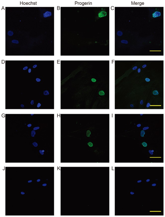
In HGPS fibroblast cultures, progerin positive nuclei are mostly lobulated or trabeculated (A–C). Human myogenic cells transfected with PMOs also demonstrated abnormally shaped progerin reactive nuclei (D–F: transfected with 1 µM PMO 421; G–I: 0.5 µM PMO 422). Untreated cells did not contain any detectable progerin positive nuclei (J–L). Magnification: 60×. Scale bar: 50 µm.