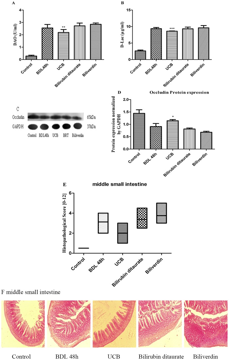
Figure 5. Effect of 0.1 mM bile pigments on the compromised gut barrier function.
After ligation of bile duct, the animals were administrated with free bilirubin, bilirubin ditaurate, or biliverdin by intragastric gavage. The serum concentrations of DAO (A) and D-Lac (B) were assayed by spectrophotometric assay after 48 h. Protein expression of occludin (65 kDa) was analyzed by Western blot (C). Occludin bands were normalized by GAPDH and expressed as relative to BDL 48 h group (D). Intestinal slices were stained with HE staining and analyzed by inverted fluorescence microscope, middle small intestine were blindly assessed for the degree of histopathology (E), all photos were captured at ×40 magnification (F). Results are mean ± SD from at least three independent experiments. *p<0.05, **p<0.01 ***p<0.001 vs. respective BDL 48 h group.

