FIG. 2.
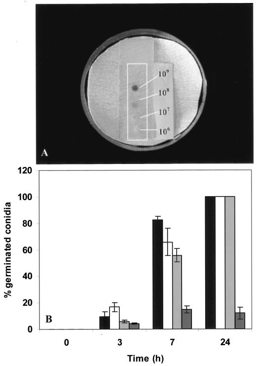
Germination efficiency at different concentrations of P. paneum conidia on MEA placed on microscope slides and harvested at 3, 7, and 24 h at conidial concentrations of 106 (black bars), 107 (white bars), 108 (light grey bars), and 109 (dark grey bars) spores ml−1. (A) Conidia suspensions on the microscopic slide after 24 h. (B) Results of the assessment of germination efficiency are shown. Three independent experiments were performed, and the error bars show the standard deviations.

