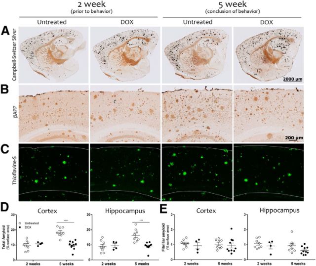
Figure 3.
Amyloid load is identical between treatment groups at the start of behavioral testing and remains stable with dox treatment. Silver staining (A), anti-Aβ immunohistochemistry (B), and thioflavine-S staining (C) were used to assess amyloid burden of the cortex and hippocampus of APP/TTA transgenic mice harvested before or after behavioral testing, following 2 weeks (2 week, left; n = 8 untreated, n = 4 dox) or 5 weeks of differential treatment, respectively (5 week, right; n = 8 untreated, n = 10 dox). D, Aβ immunostaining was used to measure total amyloid burden as a percentage of surface area in the cortex and hippocampus. In both regions, amyloid load remained steady during behavioral testing in dox-treated mice, but increased in untreated animals. The two treatment groups had similar amyloid loads at the start of testing, but were significantly different by the end (***p < 0.001). E, Thioflavine-S staining was used to measure fibrillar amyloid in the cortex and hippocampus. The area of thioflavine-S staining was identical between groups before behavioral testing, and remained unchanged over the 3 weeks of behavioral testing.

