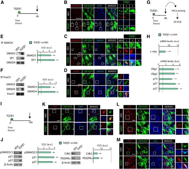
Figure 5.
TGFβ signaling modulates c-myc and p21 expression in OP cells during SCWM. A, G, I, Time line representing the paradigm used for TGFβ1 or vehicle treatment in vivo. B, Immunofluorescence analysis in the SCWM was performed after 4 h of TGFβ1 administration. Activation of TGFβ signaling induces nuclear localization of pSMAD2/3 (B, C), SMAD4 (C, D), and FoxO1 (D) in OPs. E, F, Coimmunoprecipitation analysis of SCWM extracts from TGFβ1 or vehicle (VEH)-treated mice, at 4 h after treatment. TGFβ signaling activation increased the interaction of SMAD3 and SMAD4 with FoxO1 and Sp1 in OPs in the SCWM. SCWM immune complexes were incubated with antibodies against SMAD4 (E) or FoxO1 (F). As a loading control, coimmunoprecipitation samples were blotted with the same antibodies used to perform the immunoprecipitation. G, H, P5 CNP-EGFP pups received a single administration of Veh or TGFβ1 (100 ng/kg) and 8 h later, CNP-EGFPlow (OPs) were FACS sorted to isolate RNA and perform RT-PCR analysis for genes involved in the TGFβ-mediated anti-mitotic program and OL differentiation. I, J, Proteins involved in the TGFβ-mediated anti-mitotic program and OL differentiation were analyzed by WB analysis from total extracts of SCWM after 16 h of treatment. K–M, P5 CNP-EGFP mice received TGFβ1 or vehicle, and immunohistochemistry analysis was performed in the SCWM after 16 h of treatment. Representative confocal images of proteins involved in the cell cycle in OPs (CNP-EGFP+NG2+ cells), including c-myc (K), p21 (L), and p27 (M) after 16 h of TGFβ1 treatment. Histograms express results in a.u. after normalization. n = 5 brains for each time point. **p < 0.01. Scale bars: B, 50 μm; C, D, K–M, 10 μm.

