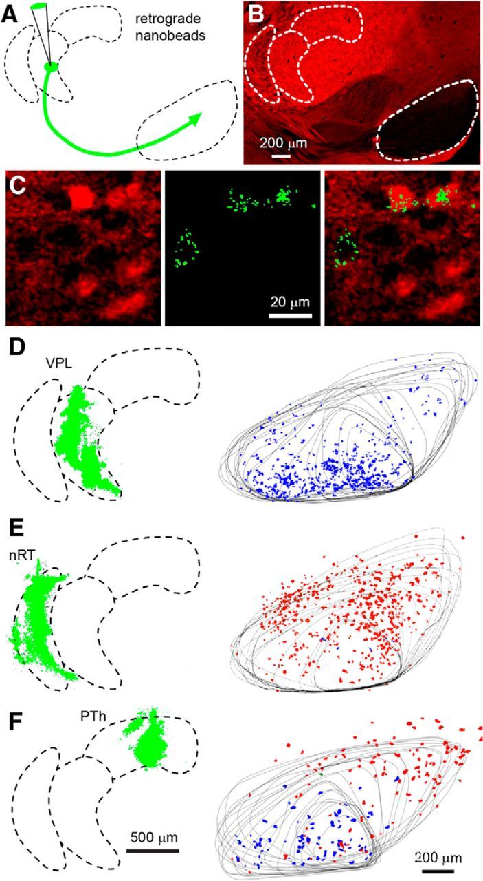
Figure 4.

Retrograde labeling of SN neurons following nanobead injection into thalamic nuclei. A, Schematic showing the injection of nanobeads into the thalamus of a vGluT2-Cre::tdTomato mouse. B, The injections were performed in vGluT2-Cre::tdTomato mice in which vGluT2-positive neurons are labeled in red. Distinct regions exhibiting differential vGluT2 density are readily identified. C, High-power image in the SN in which neurons that express vGluT2 are red (left), retrogradely labeled neurons show characteristic punctate green fluorescence (middle), and the merged image shows that, following this injection into the PTh, one non-glutamatergic and two glutamatergic neurons were labeled. D, Shows an experiment in which beads were injected into the VPL, with the injection site shown as a Z-stack determined from 17 sections (left, green). Twenty-one sections were used to determine the location of bead-labeled neurons in the SN, with the outline of the SN shown for all of the sections, and bead-labeled neurons were color coded, with red indicating vGluT2-positive neurons and blue indicating vGluT2-negative neurons. E, Example of retrograde labeling following bead injection into the nRT. F, Example of retrograde labeling following bead injection in the PTh.
