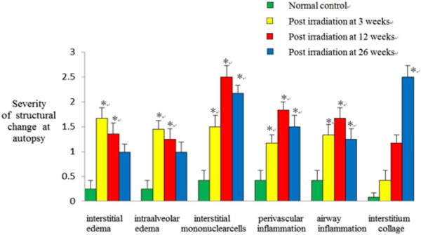
Figure 2.

Semi-quantitative assessment of morphologic changes following thoracic irradiation in autopsied specimens. For H&E staining, we evaluated an edema of the interstitial and intra-alveolar, infiltration of inflammatory cells, interstitial mononuclear cells, and interstitial collagen at each time point. Data are expressed as means ± SE (n = 8). *p < 0.05 in 2-tailed student’s t test when compared to non-irradiated controls.
