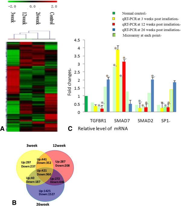
Figure 3.

Analysis of mRNA expression in the rat lung after irradiation. (A) Unsupervised hierarchical clustering of differentially expressed mRNAs in early (3w), middle (12w) and late (26w) stages of RILI. Red indicates higher expression and green indicates lower expression in irradiated lung. Black means no expression difference. The inset above represents color scales used in the cluster map. (B) Overlaps of co-regulated mRNAs (greater than a 2-fold difference) across various post-irradiation time courses. The relative levels of individual mRNA transcripts with 2-fold difference and statistical significance by Welch t-test (P < 0.05), as compared with that of a non-irradiated control rat, were identified with differentially expressed mRNAs. Data shown are real numbers of up-regulated (Up) and down-regulated (Down) mRNAs. The numbers in the overlapped areas represent the number of differentially transcribed mRNAs shared by two or three groups. (C) qRT-PCR and microarray results of mRNA (TGFBR1,SMAD7,SMAD2,SP1) expression level at each post-irradiation time point. Each reaction was performed in triplicate and the average CT was used for RQ calculation. *p < 0.05, in 2-tailed student’s t test when compared to non-irradiated controls.
