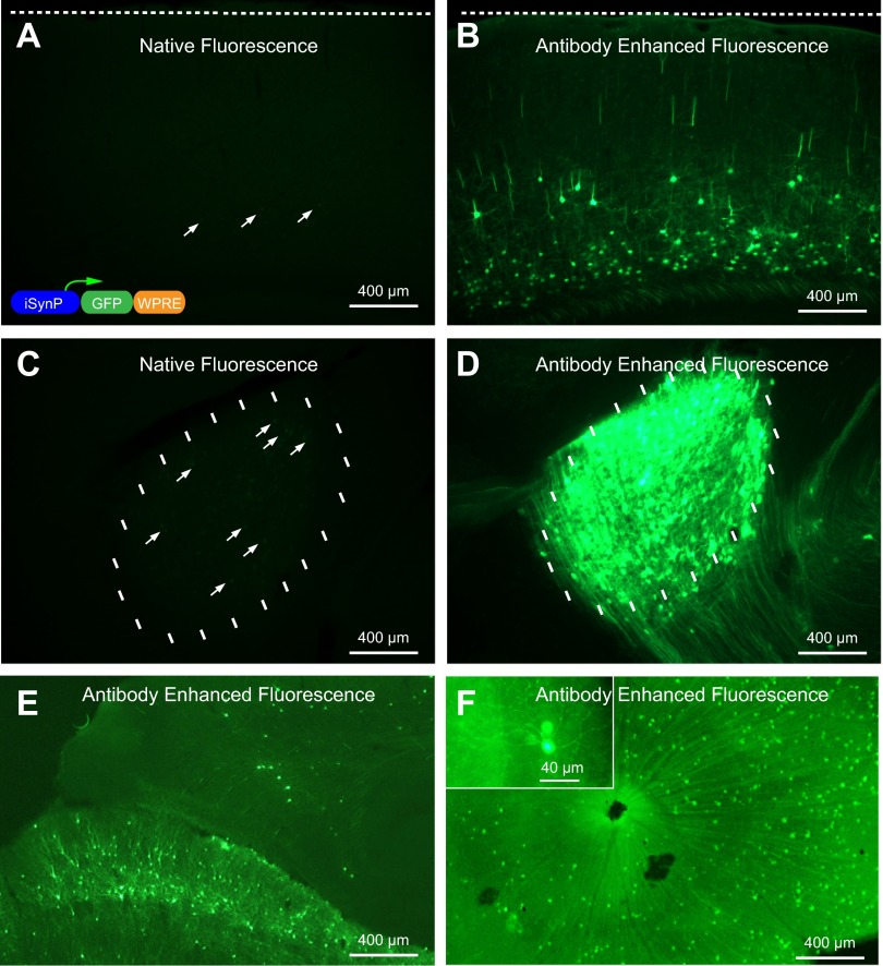
Fig. 1.
Rabies glycoprotein pseudotyping of recombinant equine infectious anemia virus (rEIAV) improved synapsin promoter (iSynP) GFP does not allow robust expression but allows immunodetection of neurons projecting to the injection site. Three weeks after virus injection into dorsolateral geniculate nucleus (DLG), the expression of GFP is barely visible under native fluorescence at the site of injection (C) and visual cortex (A), which projects to DLG. The expression can be readily detected in vast numbers of neurons after immunoenhancement with a GFP antibody at the injection site (D) and V1 (B). Other neurons labeled that are known to project to DLG include superior colliculus neurons (E) and retinal ganglion cells (F). The schematic of the viral construct is depicted in the inset in A.

