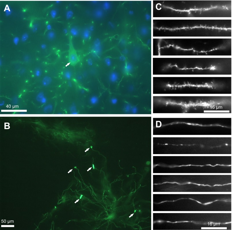
Fig. 4.
Neurons that are infected retrogradely by the TLoop rEIAV ChR2YFP virus can continue expressing the gene products as long as 6 mo. A layer V pyramidal neuron cell body is shown at high magnification with a nucleus that is indistinguishable from neighboring cells [YFP-DAPI (blue) overlay, A; cell is marked with an arrow]. Neuronal processes from many different areas from retrogradely infected neurons look normal in morphology (dendrites in C and axons in D). In rare instances, neurons that are infected by the TLoop rEIAV ChR2YFP virus that continue expressing the gene products for 6 mo show signs of fluorescent aggregations (B). Dendritic processes from neurons located in the ventral medial nucleus of the thalamus that were retrogradely infected from the barrel cortex injection and have been expressing the gene products for 6 mo show rare signs of protein Chr2YFP aggregation that may be due to uncontrolled expression (indicated with arrows).

