Fig. 1.

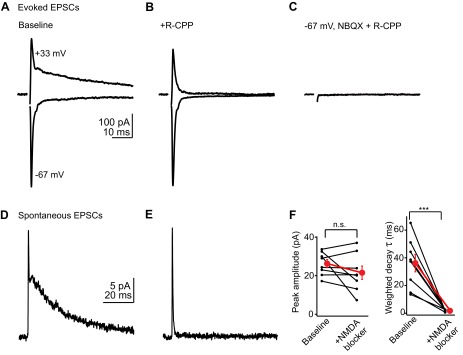
Parallel fibers activate AMPA and NMDA receptors. A: example average excitatory postsynaptic currents (EPSCs) recorded in a stellate cell at negative (bottom trace) and positive holding potentials (top trace). Of note is the slow decay component at +33 mV, typical of NMDA receptor-mediated transmission. B: in the same cell, bath application of the NMDA receptor antagonist 3-[(R)-2-carboxypiperazin-4-yl]-propyl-1-phosphonic acid (R-CPP; 5 μM) selectively blocks the slow component. C: subsequent application of the AMPA receptor blocker 2,3-dihydroxy-6-nitro-7-sulfamoyl-benzo(f)quinoxaline (NBQX; 10 μM) abolishes the fast EPSC. D: average of spontaneous EPSCs (sEPSCs) recorded at +33 mV, highlighting a slow component similar to that of evoked EPSCs. These traces are from a different cell from that shown in A–C. E: average of sEPSCs recorded at +33 mV in the presence of R-CPP, showing that the slow decay is due to NMDA receptor activation. F: quantification of 6 experiments similar to those of D and E. Left graph shows the peak amplitudes of average sEPSCs before and after NMDA receptor blockade, indicating that NMDA receptors contribute minimally to the peak. Right graph shows the effect of NMDA receptor blockers on the weighted decay time constant of sEPSCs. Black lines connect data from individual experiments; red dot is mean ± SE. ***P < 0.001; n.s., not significant.
