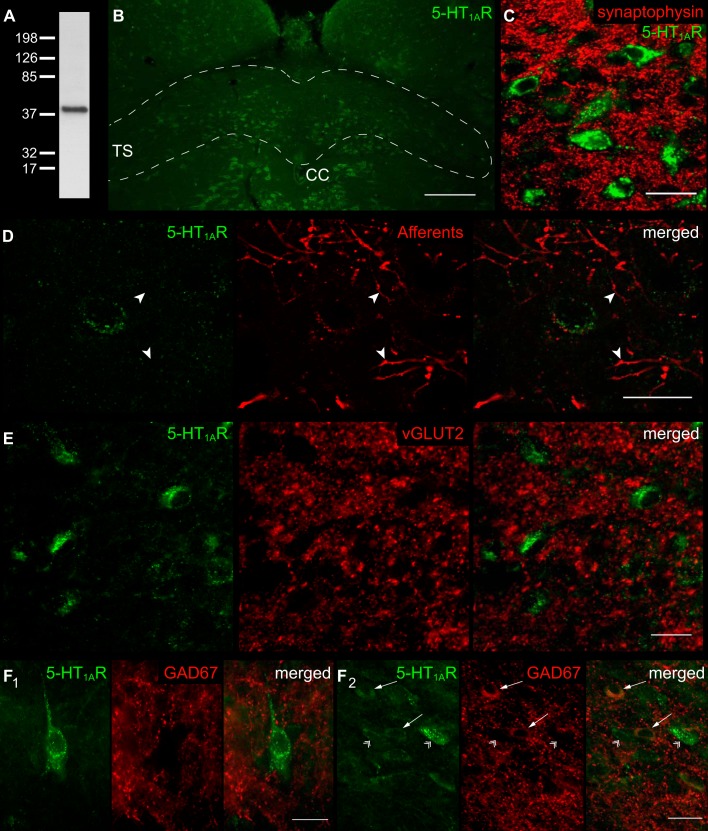
Fig. 1.
Localization of serotonin 1A receptor (5-HT1AR) within the nucleus tractus solitarii (nTS). A: immunoblot verifying antibody specificity for 50 μg of protein from the nTS. A single band for 5-HT1AR was confirmed at the appropriate size of ∼46 kDa. B: coronal section of the medial-commissural nTS (enclosed area) labeled with 5-HT1AR antibody. Scale 200 μm. TS, tractus solitarii; CC, central canal. C: nTS neurons colabeled for 5-HT1AR and synaptophysin. Note that both stainings are clearly separated, indicating that 5-HT1AR are localized somatodenritic within the nTS. Scale 25 μm. D: neuron expressing 5-HT1AR immunoreactivity with attached fluorescent Texas Red dextran labeled visceral afferent terminals. Note, afferent terminals or fibers (arrowheads) do not colabel with 5-HT1AR. The image has been background subtracted and deconvolved for clarity. Scale 20 μm. E: section showing absent colocalization of 5-HT1AR and vesicular glutamate transporter (vGLUT-2), a marker for glutamatergic terminals. Scale 20 μm. F: 5-HT1AR immunoreactivity in close apposition to glutamic acid decarboxylase (GAD67)-identified GABAergic cell terminals (F1), and colocalization with GABAergic cell bodies (F2, arrows). Note the absent colocalization of 5-HT1AR and GAD67-negative cells (arrowheads). Scale 20 μm.

