Fig. 2.
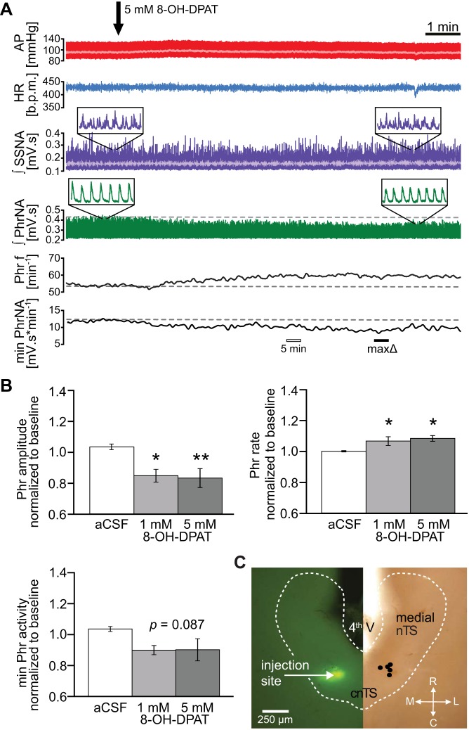
Activation of 5-HT1ARs decreases phrenic (Phr) motor output. A: representative example of pulsatile arterial pressure (AP; red), mean AP (light red trace superimposed on AP), heart rate (HR; blue), integrated splanchnic sympathetic nerve activity (∫SSNA; purple; mean SSNA in light purple) and integrated phrenic nerve activity (∫PhrNA, green). Phrenic frequency (Phr f) and minute activity (min PhrNA) are shown in the bottom panels. Note the decrease in PhrNA amplitude and increase in PhrNA rate after 8-hydroxy-2-(di-n-propylamino)tetral (8-OH-DPAT) injection (arrow), resulting in an overall decrease of minute PhrNA. Open bar depicts the point of the “5 min” measurement used for B (25-s average). Solid bar indicates time of maximum change in minute PhrNA of this individual example. B: group data (n = 5) describing the 8-OH-DPAT-induced decrease in PhrNA amplitude, increase in rate and the resulting minute nerve activity (measured 5 min after injections; open bar in A) compared with artificial cerebrospinal fluid (aCSF) control. *P ≤ 0.05; **P < 0.01 vs. aCSF. C: verification of 8-OH-DPAT injection sites. Horizontal slice of the medial-cnTS shows an example of a fluorescent bead injection (left) and the schematic summary of all injection sites (right). 4th V, fourth ventricle; bpm, beats/min; L, lateral; M, medial; R, rostral; C, caudal; cnTS, commissural nTS.

