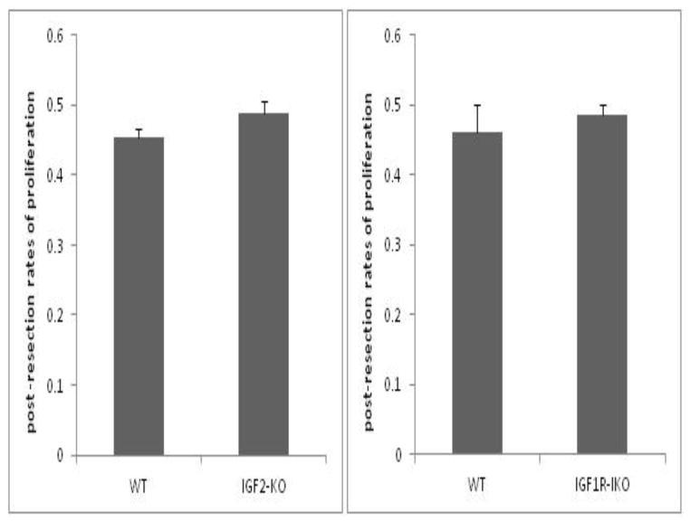
Figure 3.
Post-operative rates of crypt cell proliferation in (A) IGF2-KO and (B) IGF1R- intestinal knockout (IKO) compared to their wild type (WT) littermates. BrdU was injected 90 minutes prior to harvest. Immunostaining was performed on tissue sections and BrdU- positive stained cells were counted in each crypt. A ratio was calculated as number of cells stained positive divided by total number of cells in the crypts. A minimum of 20 crypts were counted per mouse. No statistical differences were observed between either mouse strains versus their corresponding WT controls.

