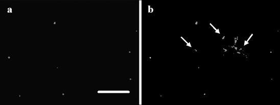
FIG. 7.
Fluorescence microscopy of a sample of aroA cells collected from a sterilized fish tank water microcosm after 70 days of incubation at 6°C. The cells were stained with the LIVE/DEAD BacLight viability kit. The images show the same field of a sample viewed for propidium iodide fluorescence (a) and for SYTO 9 fluorescence (b). The arrows in panel b indicate cells that were not stained for propidium iodide. Bar, 5 μm.

