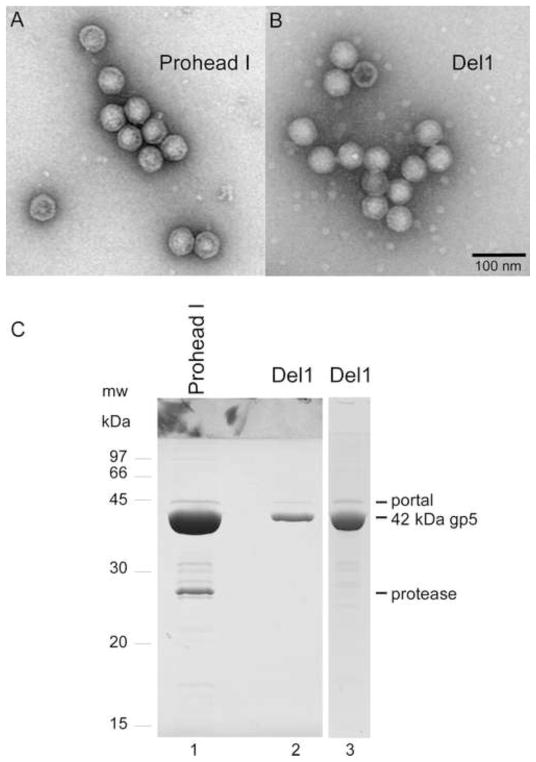
Fig. 4. Comparison of Del1 proheads (deleting residues 9–13) with wild-type proheads.
A and B. Electron micrographs of purified particles stained with 1% uranyl acetate. Both images were taken at the same magnification. The scale bar represents 100 nm. A. Wild-type Prohead I. B. Del1 proheads. C. SDS gel analysis of the composition of proheads: lane 1, Prohead I with portal and inactivated protease from plasmid pVP0-g4H65A; lanes 2 and 3, portal-containing Del1 proheads made in the presence of inactivated protease from plasmid pVP0-g4H65A-Del1.

