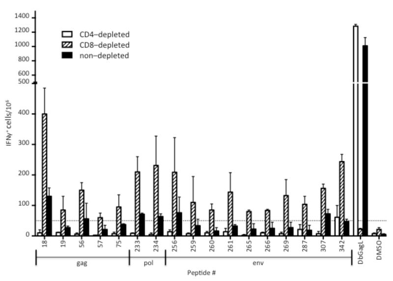
Figure 3. Reactivity of CD4- and CD8-depleted splenocytes against positive peptides.
Splenocytes from FV-infected mice at 12 dpi were depleted of either CD4+ cells (open bars), CD8+ cells (striped bars), or neither cell type (solid bars) as described in the methods. All samples were depleted of Ter119+ erythroid cells. The bar graph depicts the mean number (± s.d.) of IFN-producing cells per 106 spleen cells responding to the indicated 18mer peptide from two FV infected mice. The dashed line represents the threshold of 50 spots per 106 spleen cells. DbGagL is the positive control CD8+ T cell epitope. DMSO is the diluent control with no peptide.

