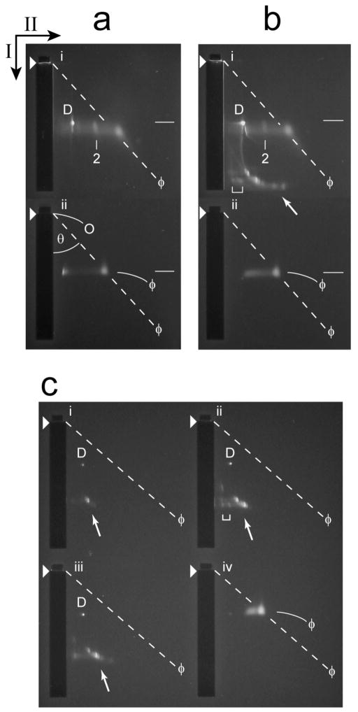
Figure 7.
2d-AGE of untrimmed heads. (a) Analysis by 2d-AGE (Materials and Methods Section; 6.0 hr. first dimension; 16.0 hr. second dimension) was performed for particles from the Head fraction of a cesium chloride density gradient like the one in Figure 3b; the growth medium had 0.1 M NaCl. The analysis was performed immediately after dialysis of the cesium chloride (panel i). Phage T3 particles were also analyzed (panel ii). (b) Analysis by 2d-AGE (6.0 hr. first dimension; 16.0 hr. second dimension) was performed two weeks after dialysis and subsequent storage at 4 °C (panel i). Phage T3 particles were also analyzed (panel ii). (c) Analysis by 2d-AGE (4.5 hr. first dimension; 14.5 hr. second dimension) was performed for the following fractions of the sucrose gradient in Figure 3b (panel, followed by fraction number, the latter as indicated in Figure 4): i, 4; ii, 5; iii, 6. Phage T3 particles were also analyzed (panel iv). Black arrows indicate the directions of electrophoresis in the first (I) and second (II) dimensions; arrowheads indicate the leading edges of sample wells. The symbol, ϕ, indicates phage bands and also the dashed, diagonal lines derived from these bands, as described below. Other symbols: D, band of protein-free DNA; O, effective origin; θ, angle between the following two lines, both originating at the effective origin: line in the direction of the first electrophoresis and the diagonal line from the effective origin through the center of a band, in this case the ϕ band in panel iv (dashed line from panel iv, which was then translated to the other panels); white arrow, indication of the position of modified heads; bracket, indication of the position of presumed dimers of modified heads; number 2, position of presumed dimers of conventional heads. As discussed in the Methods Section, RE increases as θ decreases.

