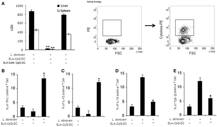
Figure 1.
Soluble leishmanial antigen–CpG–DC vaccination mediates effective protection against visceral leishmaniasis through a potent pro-inflammatory response. (A) Mice were vaccinated with SLA and CpG-ODN-pulsed DCs, SLA and control-ODN-pulsed DCs, or phosphate buffered saline (PBS; control) followed by intravenous infection with 1 × 10⁷ stationary phase Leishmania donovani promastigotes after 7 days. Mice were sacrificed on day 56 after infection. Levels of parasite burden in liver and spleen samples were determined by stamp–smear method and expressed in Leishman Donovan Units (LDU). Results are from three independent experiments and represent the mean values ± standard errors of the means for five animals per group per time point. **P < 0.001, compared to PBS-treated infected mice. In another set of experiments, splenocytes (2 × 10⁶) from control, L. donovani-infected (28 days), and SLA–CpG–DC-vaccinated infected mice (28 days) were assessed for intracellular (B) IFN-γ, (C) IL-12, (D) IL-10, or (E) TGF-β staining, which was performed as mentioned in Section “Materials and Methods” and analyzed by flow cytometry. Magnetically purified CD4+ T cells were analyzed for IL-12-PE, IFN-γ-PE, IL-10-PE, or TGF-β-PE staining to detect CD4+IL-12+, CD4+ IFN-γ+, CD4+IL-10+, or CD4+ TGF-β+ T cells. The bar graphs represent the mean dot plot values based on the region encircling positive cells from three independent experiments *P < 0.05, compared with infected sets. The error bars represent mean ± SD of three mice per group.

