Figure 5.
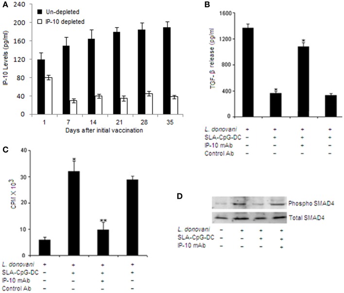
IFN-γ-inducible protein 10 depletion abrogates the TGF-β signaling in CD4+CD25+ Treg cells in SLA–CpG–DC-vaccinated parasitized mice. (A) Assessment of depletion efficiency. Levels of IP-10 were measured at indicated days from IP-10 depleted and non-depleted SLA–CpG–DC-vaccinated parasitized mice. Splenic cells were isolated at indicated time points and levels of IP-10 were measured by sandwich ELISA. The experiments were carried out twice. (B) CD4+CD25+ Treg cells (1 × 10⁶) purified from spleen of indicated groups of L. donovani-infected (28 days) mice were stimulated with SLA for 72 h. Level of TGF-β in cell culture supernatants of indicated treatment groups was determined by ELISA. Asterisks indicate statistically significant induction (*P < 0.05, **P < 0.001) of TGF-β production compared with infected sets. (C) CD4+CD25+ T regulatory cells and CD4+CD25− T cells were purified from spleen of differently treated mice (see Materials and Methods) after 28 days post-infection. CD4+CD25− T cells (5 × 10⁵) and T-depleted, mitomycin C-treated syngeneic APCs (5 × 10⁵) were stimulated with SLA (10 μg/ml) in the presence of splenic CD4+CD25+ T cells (1:1) for 4 days. Proliferation was determined by an 18-h (3H) thymidine incorporation assay. Data were presented as cpm × 103. **P < 0.001 compared with infected sets. (D) In a separate experiment, CD4+CD25+ Treg cells (2 × 10⁶), purified from spleen of indicated groups of mice, were stimulated with SLA for 30 and 60 min. The cells were lysed and subjected to Western blotting with anti-pSMAD4 and anti-SMAD4 as described in Section “Materials and Methods”. The experiments were carried out three times, of which one set of representative data is shown.

