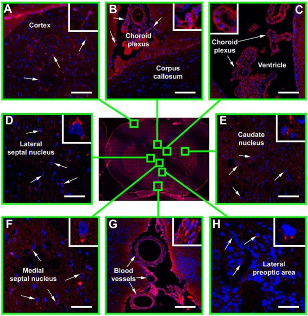
Figure 2.

Immunofluorescence confocal microscopy of Hsp70 distribution in the ischemic rat brain.
Notes: Following transient occlusion of the right middle cerebral artery (45 minutes), Hsp70 conjugated with Alexa Fluor® 555 (Invitrogen™; Thermo Fisher Scientific, Waltham, MA, USA) (red) (5.0 mg/kg) was injected into the femoral vein. After 24 hours, the animal was sacrificed and its brain was extracted and sectioned. Nuclei were additionally stained by 4′,6-diamidino-2-phenylindole (DAPI) (blue). Shown here is a brain section obtained in the coronal plane 3 mm posterior to the bregma (stereotaxic atlas of Pelligrino, 1979). The presence of Hsp70 was assessed in the following structures: (A) brain cortex; (B) corpus callosum; (C) ventricle; (D) lateral septal nucleus; (E) caudate nucleus; (F) medial septal nucleus; (G) blood vessels; (H) lateral preoptic area. Scale bar: 25 μm.
