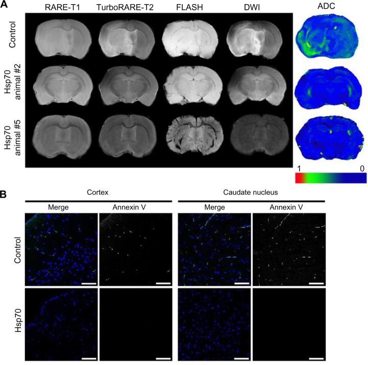
Figure 5.
Assessment of the infarcted zone following treatment with Hsp70-loaded alginate granules.
Notes: (A) Representative magnetic resonance images of rat brains from the control and Hsp70-treated groups (animals #2 and #5). Magnetic resonance imaging was performed according to the RARE-T1, TurboRARE-T2, FLASH, and diffusion-weighted imaging (DWI) regimens. Additionally, DWIs were evaluated using apparent diffusion coefficient (ADC) maps. (B) Confocal microscopic images of brain sections were obtained in the coronal plane. Apoptotic cells were detected using an annexin V kit (green). Nuclei were stained with 4′,6-diamidino-2-phenylindole (DAPI) (blue). Scale bar: 25 μm.

