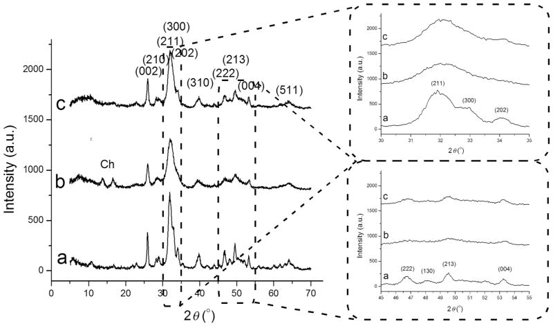
Figure 2.
X-ray diffractograms of HAp (a), HAp/chitosan (b), and clindamycin-loaded HAp (c) powders synthesized in this study, along with {hkl}indices ascribed to the most intensive reflections of crystalline HAp. Diffraction peaks originating from chitosan are labeled with “Ch.” The two inlets show magnified 2θ = 30°–35° and 45°–55° regions.

