FIGURE 1.
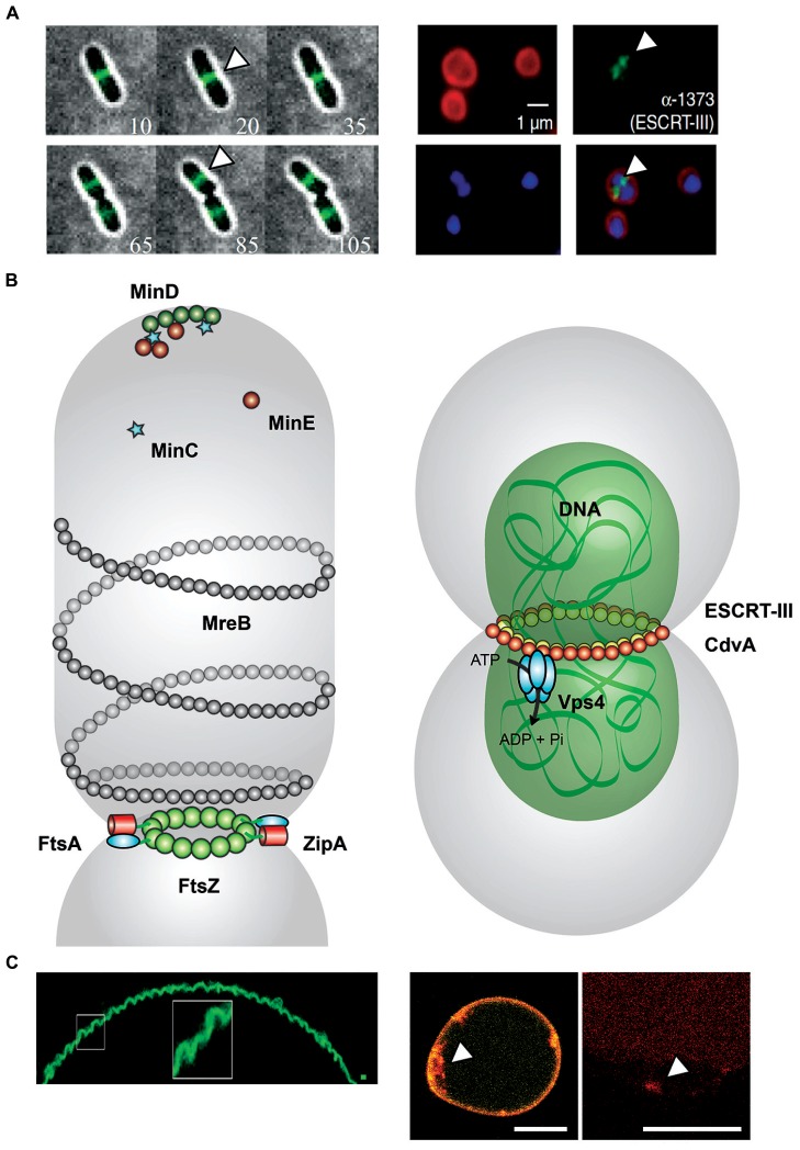
Comparison between FtsZ- and ESCRT-III mediated cell division. (A) Left: Montages from time-lapse movies of Escherichia coli cells expressing FtsZ–GFP during cell division. At each time point, the fluorescence image (green) was overlaid with the corresponding bright-field image (gray) and the time (in minutes) displayed in the bottom corner. FtsZ (white arrowheads) forms a clear band at mid-cell early and remains there throughout the division process (Buss et al., 2013). Right: Localization of ESCRT-III in Sulfolobus acidocaldarius. Representative images show the FM4-64X staining for membrane (red), DAPI staining for DNA (blue), antibody labeling of ESCRT-III, and merged images. ESCRT-III localization is visualized by white arrowheads. Scale bar, 1 μm (Samson et al., 2008). (B) Left: FtsZ-mediated cell division in Escherichia coli. During the initial stage of proto-ring assembly, FtsZ (green) is located to mid-cell by the oscillating Min waves [MinD (dark green), MinE (dark red), and MinC (light blue stars)]. FtsZ attaches to the membrane-associated FtsA (cyan). ZipA (red) binds competitive with FtsA to FtsZ. ZipA–FtsZ binding also increase membrane fluidity (Rico et al., 2013). MreB (gray) structures colocalize with Z ring at mid-cell (Fenton and Gerdes, 2013). Right: ESCRT-III-mediated cell division in Sulfolobus acidocaldarius. CdvA (Saci_1374, yellow) and DNA (green) build up double-helical structures horizontally to the cytokinesis region in Sulfolobus acidocaldarius (gray). ESCRT-III (Saci_1373, red) and paralogs interact with CdvA via its C-terminal winged Helix-like domain and also bind to MIT domain of the Vps4 (Saci_1372, cyan). The AAA-type ATPase Vps4 regenerates the ESCRT-III complex after cytokinesis (Moriscot et al., 2011; Samson et al., 2011). (C) Left: Membrane curvature induced in GUVs by assembly of FtsZ filaments. Under low membrane tension conditions, MTS-FtsZ-YFP showed spontaneous deformation of the GUV membranes (Arumugam et al., 2012). Right: Membrane curvature events in GUVs by the ESCRT-III machinery. By droplet emulsion transfer method CdvA, ESCRT-III (red Alexa647 labeled) and Vps4 were inserted into DOPC GUVs (yellow). The location of ESCRT-III (white arrowhead) to curved membrane suggests a function in membrane deformation event. Scale bar, 10 μm.

