FIGURE 2.
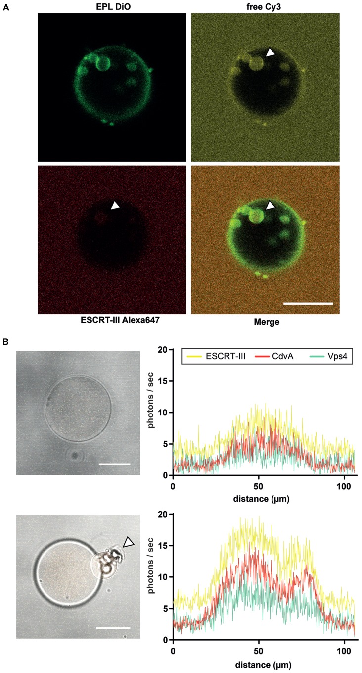
Liposome preparation for intraluminal experiments. (A) ILV formation into EPL (Escherichia coli polar lipids) GUVs in presence of ESCRT-III and Vps4. The GUV membrane is stained green with DiO. Red labeled ESCRT-III proteins (Alexa647), unstained Vps4 proteins and free Cy3 were added to the extraluminal buffer of the EPL GUVs. After ILV formation of the GUV membrane ESCRT-III as well as the soluble marker Cy3 could be visualized in the vesicles (white arrowheads). Scale bar, 10 μm. (B) De novo protein production in GUVs via protein synthesis kit within 3 h. Left figures reveal brightfield images of a GUV with recombinant CdvA-mCherry, Vps4-AmCyan1 and ESCRT-III-zYellow1 plasmids in combination with a protein synthesis kit included. The low fluorescence level (histogram in upper right) of all three proteins indicates the beginning of de novo production. After 3 h of incubation the fluorescence of all proteins are increased (histogram in lower right) and it could be observed a budding event at GUV (lower left image, white arrowhead). Scale bar, 10 μm.

